Magnetoencephalography (MEG) is a non-invasive brain imaging technology that can identify the areas of the brain where seizures originate and the areas of the brain that control language, sensation, motion and vision by creating real-time pictures of your child’s brain activity. Identifying these locations can help doctors understand how these functions have been affected by underlying conditions like epilepsy or brain tumors and, if needed, how to plan for surgery.
MEG is used in conjunction with other imaging technologies like MRI, which document the structure of the brain, to provide a complete picture of the brain’s functional architecture giving the most accurate view of your child’s condition.
Using hundreds of sensors, MEG detects the small magnetic fields created by electrical signals produced when brain cells are active. MEG can detect the time (within milliseconds) and location (within millimeters) of these signals to create an image of brain activity unfolding in real-time with a high degree of accuracy.
To learn more about Le Bonheur’s MEG or to schedule an appointment, call (901) 287-7130.
Benefits of a magnetoencephalography (MEG) scan
In addition to being non-invasive, MEG has several advantages.
- Safe and radiation-free
- Easily repeatable with no time limit to the scan
- Able to have visual, auditory, tactile and motor contact with the environment during the scan
- Brain activation can be mapped within milliseconds and millimeters of accuracy
- Proven to be as effective as invasive brain mapping techniques such as electrocorticography and Wada procedure
- Able to used under sedation when necessary and still map epilepsy activity and the functional areas of the brain
MEG scans are beneficial for patients with epilepsy to:
- Locate where seizures occur in the brain
- Map the reorganization in the brain due to epilepsy, tumor or other change in brain structure
- Determine if surgery is a viable option for the patient
MEG scans are used prior to brain surgery in order to:
- Completely and accurately map brain function to best preserve it after surgery
- Dictate surgery options and create a surgery plan
- Predict consequences to functionality of the brain after surgery
Why Le Bonheur’s MEG?
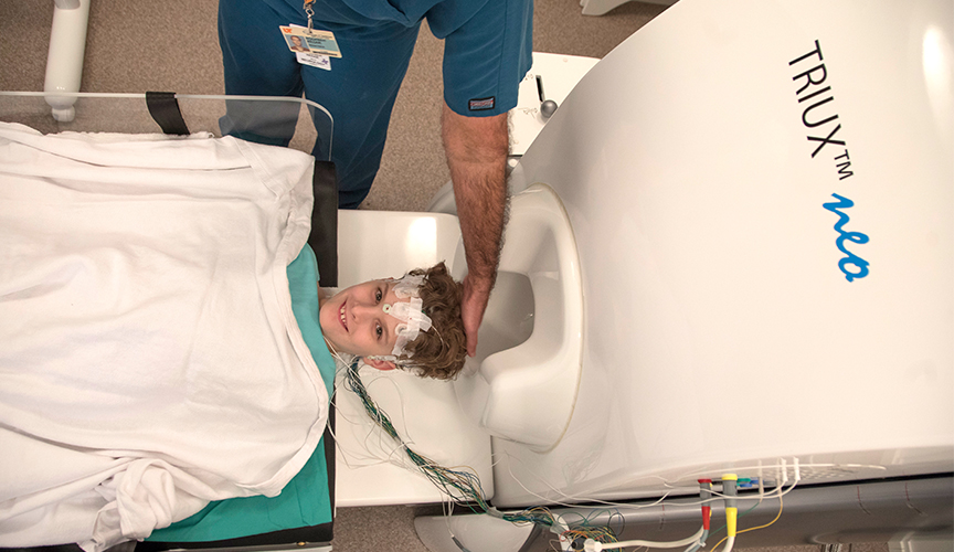
Le Bonheur uses the most up-to-date MEG technology, TRIUX neo MEG – a new brain scanning technology for functional brain imaging. We were the first hospital in the world to install this next-generation magnetoencephalography (MEG) technology.
Le Bonheur’s MEG instrument and program are unique in several ways:
- Sedation is available tailored to each child’s age and need while still maintaining the effectiveness of the test.
- The latest MEG technology allows us to scan children with VNS or other implanted medical devices who previously could not undergo MEG scans.
- We have a dedicated neurology child life specialist who prepares children in advance of the MEG scan and provides distraction and engagement when needed.
- FedExFamilyHouse, a 75-room and-suite hotel, is available for out-of-town patients free of charge.
Undergoing a MEG scan
- To eliminate noise and detect the small signals coming from the brain, the MEG is housed in a special room designed to provide the ideal environment for testing.
- The part of the MEG which records the brain activity is known as the helmet and contains several hundred sensors to cover the surface of the head. MEG does not create any noise when it is operating, and the helmet leaves your child’s face uncovered for the duration of the scan.
- A two-way intercom and camera are available so that the patient and MEG technician can communicate with each other during the scan
- Breaks can be taken during testing if your child needs to move.
- Upon arrival to the MEG suite, your child will have to remove any jewelry, accessories or other metallic objects, and we’ll verify if your child has any implanted medical devices. In some cases, your child may need to change into a hospital gown or scrubs.
- You and your child will then proceed to the room that holds the MEG and be given an explanation of how the machine works.
- During this time, your child’s head will be placed inside the helmet for a brief (around two minutes) recording of their brain activity.
- This first scan helps the testing staff identify anything that might interfere with the MEG recording and familiarize the patient with the testing environment and what to expect throughout the duration of the procedure.
- We will also record an EEG as your child undergoes the MEG scan to help interpret the testing results. Our technicians will place about 20 EEG electrodes on your child’s head while they are laying down on a soft bed outside the testing room.
- The technician will prep your child’s head with a cotton swab and gel to clean the skin and then place the EEG electrodes on the head using paste to ensure a good contact.
- To accurately match brain activity recorded by the MEG to the patient’s MRI, our technicians map the shape of your child’s head and to track head location inside the MEG helmet throughout the testing procedure.
- Your child will be seated in the prep area and put on a pair of plastic glasses.
- Five small coils that look like sequins will be taped to the forehead and behind the ears, and three small sponge donut-shaped stickers will be taped above the nose and in front of the ears.
- Our technician will map the locations of these coils and stickers by gently touching them using a plastic pen.
- Using the same plastic pen, the technician will make an outline of the patient’s head by gently running the tip of the pen over the surface of the scalp, forehead, nose and cheeks.
- The amount of time for the scan depends on a wide variety of factors, but usually does not exceed three hours, including preparation and break periods in the scan.
- The MEG scan can be broken down into two phases – epilepsy mapping and functional mapping – to identify areas of the brain that are for these functions.
- Epilepsy mapping records your child’s brain activity while in a restful, still state for about 45-60 minutes with the goal of capturing interictal events – abnormal patterns of brain activity that occur in between seizures.
- Functional mapping identifies where basic functions such as language, sensation, movement and vision are located inside your child’s brain and how they might be affected by seizure areas or a tumor.
- The language mapping test is performed by the MEG technician saying a series of words, through ear phones, and mapping the brain’s response. The test lasts about 20 minutes total.
- The sensory mapping test helps identify where sensation in the hands is located in your child’s brain. A small, painless electrical signal is given to the wrist to make the thumb twitch. This test last about 12 minutes with both the left and right hands.
- Our neuroscientists will analyze, interpret and compile the results of the MEG scan in a comprehensive report shared with your physician.
- Your physician will discuss the results with you prior to discharge from the hospital or at your child’s next clinic appointment.