Brain Blueprint
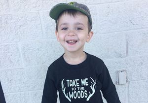
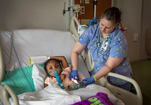
Bree Sudduth of Fayetteville, Ark., was desperate for answers for her son, Tristan.
Diagnosed at birth with Tuberous Sclerosis Complex, Tristan, now 3, had experienced seizures since the first few months of his life. After visiting multiple neurologists and emergency rooms, Bree and Tristan arrived at Le Bonheur Children’s Neuroscience Institute looking for relief.
“Tristan was delayed in every area,” said Bree. “He had an 18 month level of speech at 2 years old, he had to wear a helmet at all times and he couldn’t even run.”
Physicians got to work on finding where his seizures originated – looking first to a suite of functional brain imaging modalities that give them a leg up on seizure disorders.
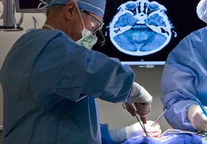
The newest of that technology, the MEGIN’s TRIUX™ neo MEG (magnetoencephalography) system, keeps neuroscientists on the cutting edge of finding answers for epilepsy and brain tumors. Le Bonheur Children’s is the first hospital in the world to install the new MEG system. Staying on the forefront of mapping technology is key to the program’s success, said Chief Pediatric Neurologist James Wheless, MD, who also serves as co-director of the Neuroscience Institute.
We know this has the capability to really benefit children and their families,” Wheless said. “If we can do that early on, we can change their entire lives.
A CUTTING-EDGE TECHNOLOGY
TRIUX™ neo MEG provides the capability to map the brain’s language, sensory and movement centers with 248 sensors that non-invasively detect the miniscule magnetic fields created by electrical signaling in neurons. MEG recordings can subsequently be superimposed over the patient’s CT or structural MRI scan to provide a complete picture of the brain’s functional architecture in real-time.
The clinical utility of MEG is highlighted by Roozbeh Rezaie, PhD, and Abbas Babajani-Feremi, PhD, the two principal neurodiagnostic team members applying MEG to assist in the treatment of epilepsy and brain tumors.
“MEG imaging works by reconstructing activation inside the brain based on magnetic fields outside of the head that are associated with the electrical activity that goes on in neurons,” says Rezaie, Director of Le Bonheur’s Magnetoencephalography (MEG) Laboratory. “MEG often completes the picture and provides a complementary approach to other imaging modalities to provide the surgeon with an accurate picture of the brain areas responsible for language, sensory and motor functions.”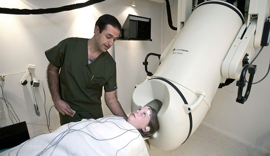
Last year many Le Bonheur patients could not undergo MEG testing because of their vagus nerve stimulation (VNS) implant. VNS is a treatment option for patients with intractable epilepsy that works similar to a pacemaker, sending mild electric pulses to the vagus nerve to control seizures. Previously, MEG could be used for mapping prior to VNS implantation, but once VNS was installed, the patient could no longer undergo a MEG scan due to the extreme noise generated by the presence of a foreign metal object inside the patient’s body.
With the advanced technology of the TRIUX™ neo MEG system, patients can undergo MEG before and after VNS implantation to better measure the efficacy of the device in controlling seizures.
Currently Babajani-Feremi is studying whether MEG can predict which patients will respond well to VNS prior to implant – a question that has previously been impossible to forecast.
“VNS is a low-risk surgical option for patients with drug resistant epilepsy,” said Babajani-Feremi. “Our results showed there are biomarkers that can be detected in a MEG scan to identify patients who would benefit from VNS.”
A SURGICAL DECISION
For children with epilepsy or brain tumors, Le Bonheur’s imaging modalities for mapping the brain play a crucial role in determining whether surgery is a good option for a patient.
Even when surgery is the best choice, it still carries the risk of removing or injuring a part of the brain that controls functionality – whether it is motor, sensory or language. Brain imaging helps to dictate the options for surgery and predict consequences to functionality of the brain after surgery.
“The different imaging modalities come together to show how aggressive we can be with our brain surgery,” says Frederick Boop, MD, co-director of the Neuroscience Institute and chairman of the Department of Neurosurgery for the University of Tennessee Health Science Center. “For example, if a tumor is near a speech area, we use imaging to prevent injury to that patient’s speech area to preserve speech post-surgery.”
Epilepsy and brain tumors add another level of complexity to brain imaging. The beauty of the brain is its flexibility and ability to restructure its pathways and reorganize the functional areas, said Shalini Narayana, MBBS, PhD, director transcranial magnetic stimulation (TMS) laboratory.
“With imaging we can find the reorganization of the brain due to epilepsy,” Narayana said. The TRIUX™ neo MEG is only one piece of the imaging puzzle. In addition to structural imaging such as MRI and CT, the functional imaging suite is conducted across disciplines. Functional MRI (fMRI) is run by the radiology department while magnetoencephalography (MEG), transcranial magnetic stimulation (TMS), high-density electroencephalogram (hd-EEG) and high gamma electrocorticography (hgECoG) are run by the pediatric neurology department. All of these modalities work in different ways to provide mapping information about brain function.
Using multiple imaging modalities for surgery is crucial to confirm that mapping is correct — as the adage says, “measure twice, cut once.” The types of imaging that the Neuroscience Institute uses for a patient depend on a variety of factors including age, cognitive ability as well as what functionality needs to be localized.
TALENT AND TEAMWORK
Brain imaging technology is only as effective as the people who operate it and interpret the results. It’s a big reason why Institute Co-directors Wheless and Boop insisted on recruiting a depth of expertise across the board to improve patient outcomes.
“Most importantly, we have the talent to use the equipment to its maximum potential,” says Boop. “This group has been working together for more than a decade and has more experience than anyone in the country.”
Le Bonheur’s three imaging experts in pediatric neurology — Babajani-Feremi, Narayana and Rezaie — each have their area of expertise.
“We have the manpower here for fast turnaround of brain imaging results,” says Rezaie. “We have access to all resources under one roof. Le Bonheur is a one stop shop with all imaging modalities along one hallway.”
After meeting with a Le Bonheur patient, the neurosurgeon or epileptologist orders imaging tests to be performed. Depending on the test needed, one of the neuroimaging experts will conduct the brain mapping.
These results are then conveyed directly to the epileptologist and neurosurgeon in order to make decisions regarding treatment or surgery and to talk to families about potential outcomes. Imaging even follows the patient into the operating room where they are integrated in real time onto the intraoperative MRI (iMRI) in the operating room.
“Dr. Boop and Dr. Wheless don’t just treat the brain imaging modalities as gadgets,” says Babajani-Feremi. “They believe in this technology, that it has immense value and can be used in operating and decision making.”
EARLY INTERVENTION
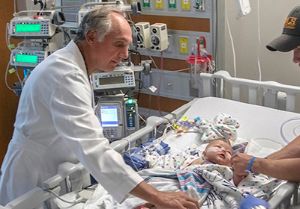
Because of this technology and teamwork, younger patients like Tristan Sudduth are able to have surgical interventions that were not previously possible.
For Tristan, neurologists used hgECoG and MEG to map his brain prior to surgery. MEG was used to identify areas where the interictal epileptiform discharges were localized.
“However, due to his age, MEG, fMRI and TMS were unsuccessful in localizing his language areas,” says Babajani-Feremi. “But with hgECoG, Tristan could sit in his mother’s lap and perform a simple object-naming task allowing us to successfully localize language areas.”
For Tristan, using these complementary imaging modalities made all the difference. He showed no language deficit after surgery or at his one-month follow-up. His mom, Bree, reports that he’s now talking in five to six word sentences.
Bree is thrilled that she can count her son Tristan as one of the successful outcomes from Le Bonheur’s Neuroscience Institute.
“I have as close to a normal child as I’ve ever had, and I can’t believe this is the same kid,” says Bree. “I want to thank all of the doctors and nurses at Le Bonheur. They saved my son.”
Help us provide the best care for kids.
Le Bonheur Children's Hospital depends on the generosity of friends like you to help us serve 250,000 children each year, regardless of their family’s ability to pay. Every gift helps us improve the lives of children.
Donate Now