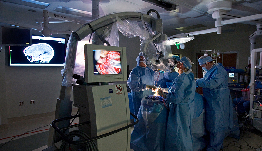
Intraoperative Magnetic Resonance Imaging (iMRI) is one of the neuroimaging technologies we use to care for children who need brain surgery.
Located in our surgery suite, the iMRI is used for pre-surgical mapping and during brain surgery. It provides our doctors and nurses with the highest quality structural image of your child’s brain. We are one of the first children’s hospitals in the country with a 3T intraoperative MRI.
Le Bonheur Children’s Hospital is proud to be the only hospital in the region equipped with an intraoperative MRI (iMRI) specifically designed for pediatric neurosurgery. This advanced imaging technology allows surgeons to capture real-time, high-resolution scans during brain procedures, enhancing precision and safety. No other hospital in or near Mississippi or Arkansas offers this level of intraoperative imaging, making Le Bonheur a regional leader in pediatric neuroscience and surgical innovation.
The Intraoperative Magnetic Resonance Imaging is an important tool in brain tumor removal surgeries. Surgeons are able to collect scans without moving the patient from the surgical table during surgery, ensuring that the optimum position is maintained. This also eliminates the need, in many cases, for additional sedation or additional surgeries.
iMRI is also used for functional testing in patients who are being considered for surgery. The information the iMRI provides complements the Neuroscience Institute’s other advanced diagnostic tools – magnetoencephalography (MEG) and transcranial magnetic stimulation (TMS). When used together, these technologies allow our experts to create a plan of care specific for your child.